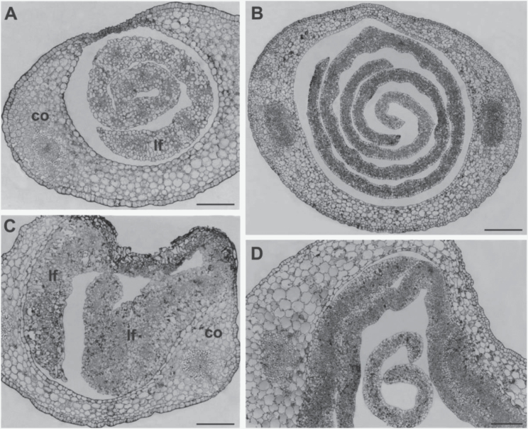
Fig. 4.
Light microscope micrographs of thin transversal sections of apical (A, C) and basal (B, D) regions of germinating seedling. (A) The apical region of a wild-type shoot shows a well-organized leaf in the hollow of the still closed coleoptile. (B) Young leaves independently enrolled are inside the coleoptile in the basal region of the wild-type shoot. (C) In the apical region of the mutant shoot masses of disorganized leaf tissues are fused with the coleoptile. (D) Fused areas between coleoptile and leaf surfaces are visible in the basal region of the mutant shoot. A young free leaf is also present. co, coleoptile, lf, leaf. Bars: 100 μm (A, C, D); 200 μm (B).

