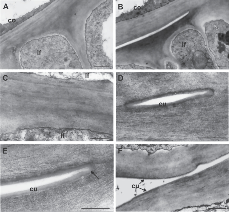
Fig. 6.
Transmission electron microscope micrographs of epidermal cell walls of coleoptile and first two leaves from the fdl1-1 mutant seedlings. (A) The cell wall proper of coleoptile and first leaf cannot be distinguished in a fused zone. (B) Distinct cell walls are visible in the free area adjacent to the fused one. (C) The higher magnification reveals the absence of any cuticular materials between the fused cell walls of two leaf surfaces. (D) A cuticle layer can be seen on the cell walls of the small free zone. (E) An evident continuous cuticle covers the free surfaces of the epidermal cells, also bordering the point of fusion (arrow). (F) The cuticle overlays the surfaces of two unfused leaf epidermises. co, coleoptile; cu, cuticle; lf, leaf. Bars: 1 μm (A, B); 0.5 μm (C, F); 0.25 μm (D, E).

