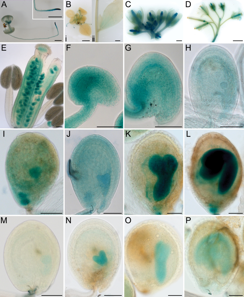
Fig. 3.
HES:GUS activity during plant development. Plant tissues were stained overnight unless otherwise indicated. (A) A 1-week-old seedling with GUS activity in the root tip, emerging leaves, and vein of cotyledons. Insert showing an enlarged root tip. (Bi) A 2-week-old seedling with GUS activity in the root tip (4h staining in A, Bi and M-P). (Bii) Stem and a cauline leaf. (C) GUS staining of upper part of the inflorescence showing early buds stained in blue. The petals, sepals, and anthers of older buds were negative for staining. (D) Lower part of the same inflorescence, showing siliques were negative for staining after development. (E) Closer image of a bud from C. (F) An unfertilized ovule just prior to pollination. (G) An early seed (~24 HAP). (H) A seed at mid globular stage. (I) A seed at late globular stage. (J) A seed at heart stage. (K) A seed at torpedo stage. (L) A seed at curled cotyledon stage. (M) A seed at late globular stage, showing GUS in the embryo and possibly in the endosperm. (N) A seed at mid heart stage, showing GUS in the embryo and weakly in the endosperm. (O) A seed at torpedo stage showing GUS activity in the embryo. (P) A seed with bent embryo ~8 day post fertilization. Scale bars: 1mm in A, C and D; 2mm in B; 100 µm in E-P.

