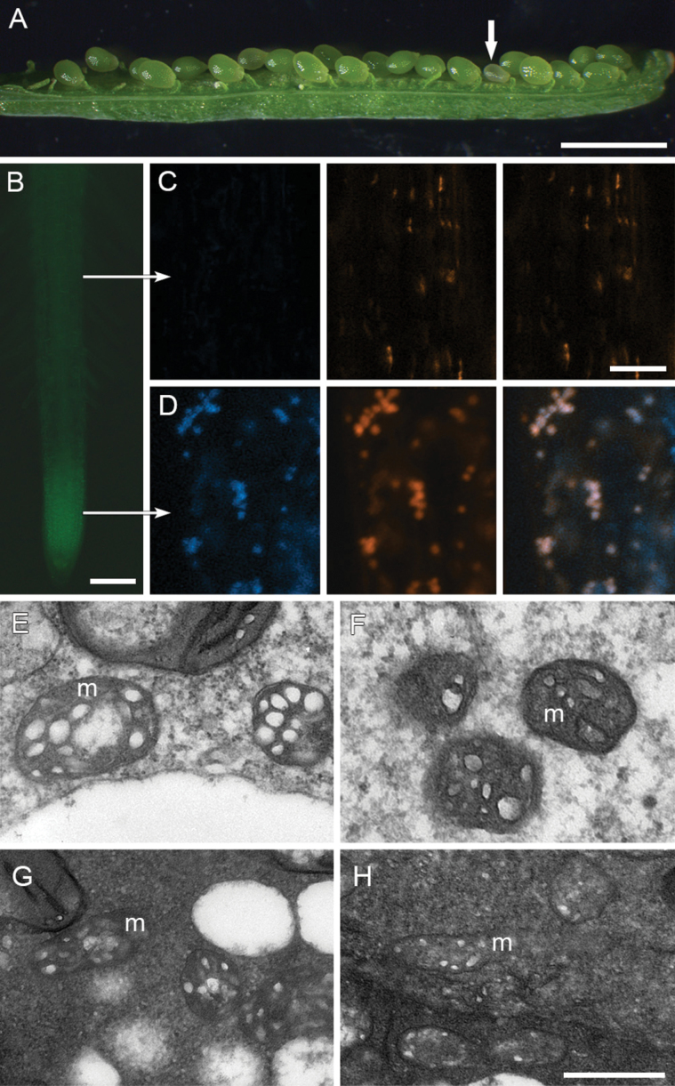
Fig. 5.
HES:GFP targets mitochondrion. (A) Silique of a hes heterozygous plant carrying HES:GFP, showing one small seed (arrow). (B) HES:GFP fluorescence in root tip. (C) Confocal microscope images in a cell from root mature region (upper arrow in B): left panel, negative HES:GFP fluorescence; middle panel, bodies stained with MitoTracker Deep Red; right panel, overlay. (D) Confocal microscope images in a cell from root tip (lower arrow in B): left panel, positive HES:GFP fluorescence; middle panel, bodies stained with MitoTracker Deep Red; right panel, overlay. (E-H) TEM images of mitochondria (m) from wild-type endosperm (E), hes endosperm (F), wild-type embryo (G) and hes embryo (H). Scale bars: 1.5mm (A); 100 μm (B); 500nm (E-H, bar shown in H), and 4 μm (C-D, bar shown in D).

