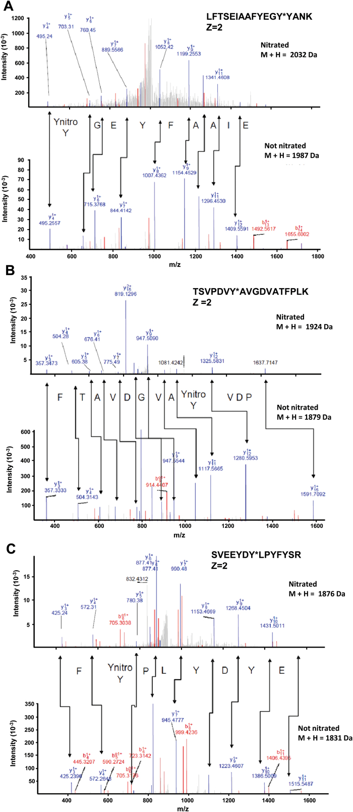
Fig. 2.
Comparison of the nitrated (top) and unmodified (bottom) MS/MS spectra of the peptides identified from the pea peroxisomal MDAR in the corresponding panels: (A) LFTSEIAAFYEGY*YANK, (B) TSVPDVY*AVGDV ATFPLK, and (C) SVEEYDY*LPYFYSR. Peptide fragment ions are indicated by ‘b’ if the charge is retained on the N-terminus and by ‘y’ if the charge is maintained on the C-terminus. The subscript indicates the number of amino acid residues in the fragment studied from either the N-terminus or the C-terminus. The superscript indicates the charge (1+ or 2+) of the backbone fragmentation. (This figure is available in colour at JXB online.)

