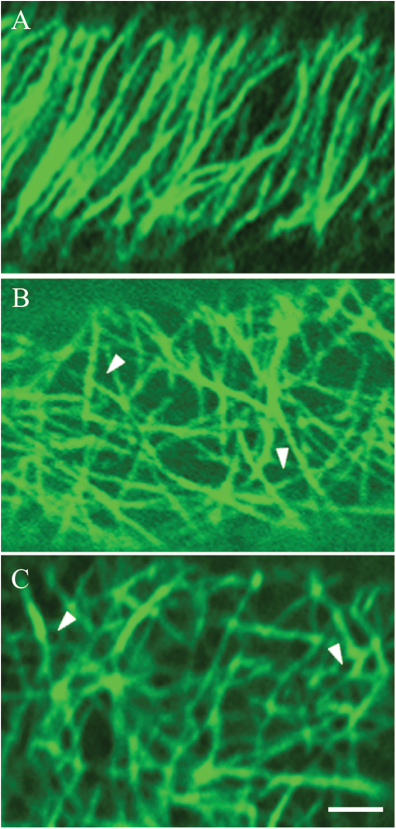
Fig. 2.
Confocal images of three categories of CMT arrays observed in adaxial epidermal cells of V. faba cotyledons cultured for 24h. CMT arrays immunolabelled with anti-α-tubulin and IgG–Alexa Fluor 488 conjugate. (A) ‘Organized’: parallel arrays of thick CMT bundles. (B) ‘Randomized’: an array defined by thick, strongly labelled CMT bundles arranged in a random network with distinctive polygonal gaps (arrowheads) in the network. (C) ‘Randomized with depletion zones’: an array composed of circular depletion zones surrounded by a possible combination of fine fragmented CMTs and tubulin monomers sometimes appearing like a collar (arrowheads). Bar, 2.5 μm.

