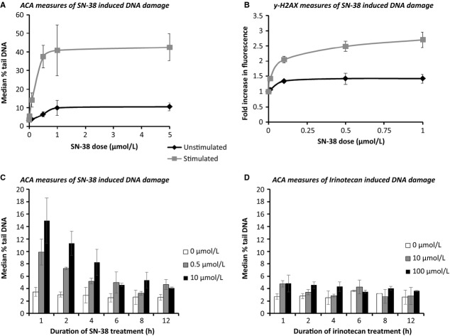
Figure 4.
Optimization of the ex vivo study assays. DNA damage measured using (A) ACA and (B) ƴ-H2AX detection in PBLs cultured in the presence or absence of a mitogen, prior to treatment with SN-38 for 1 h and DNA damage detected over a 12-h time course in PBLs cultured with PHA stimulation for 72 h prior to treatment with (C) irinotecan and (D) SN-38.

