
Figure 4.
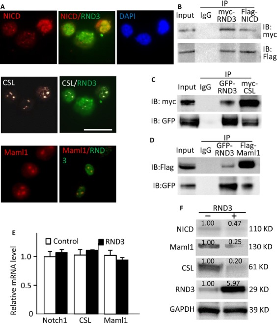
RND3 physically interacted with NICD-CSL-MAML1 complex and facilitated its degradation. (A) Immunofluorescent staining displayed the colocalization of endogenous RND3 with NICD (upper panel), CSL (middle panel), and MAML1 (lower panel) in U251 glioblastoma cells. (B-D) Coimmunoprecipitation (IP) pull-downs were performed, followed by immunoblotting analyses (IB). The blots confirmed the interactions of RND3 with NICD, CSL, and MAML1. (E) qPCR analysis indicated no changes in NOTCH1, CSL, and MAML1 transcript levels by forced expression of RND3. The data were pooled by three experiments, with analysis for each in triplicate. (F) Immunoblotting analyses showed that forced expression of RND3 resulted in decreases in NICD, MAML1 and CSL protein levels in U251 glioblastoma cells. The number at the top of each band represents the average of densitometries from three experiments, normalized by GAPDH. Scale bar represents 20 μm.