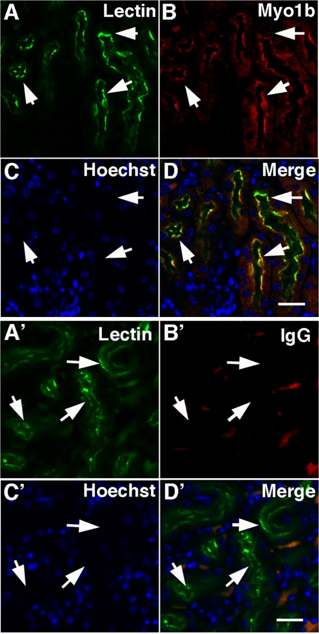
Fig 1. In mouse kidney Myo1b is found in PT cells.

In kidney cross-sections treated with Hoechst stain to identify nuclei (C, blue), Myo1b stained with anti-Myo1b antibodies (B, red) co-localized with Lotus tetragonolobus lectin, a marker for PTs (A, green). D is a merged image of A-C. The arrows identify the corresponding region in each panel. Scale bar = 20 μm. A’-D’ are control images of sections stained with lectin (A’), isotype antibodies (B’), and Hoechst (C’); D’ is a merge of A’-C’. The arrows, which identify PTs in A’, point to the corresponding location in each panel. Scale bar = 20 μm.
