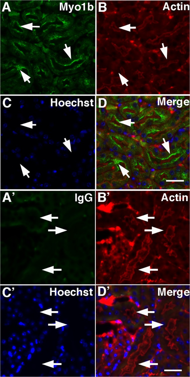
Fig 2. Myo1b localizes to the brush border of renal PT cells.

In mouse kidney cross-sections, Myo1b (A, green) identified with anti-Myo1b antibodies was at the brush border of PT cells identified by staining of apical microvilli with anti-actin antibodies (B, red). C, Hoechst stain. D, Merge. Scale bar = 20 μm. A’–D’ are control images of renal PT cells using rabbit IgG (A’). B’ is stained with anti-actin antibodies. C’ is stained with Hoechst, and D’ is a merged image. The arrows point to the corresponding location in each image. Scale bar = 20 μm.
