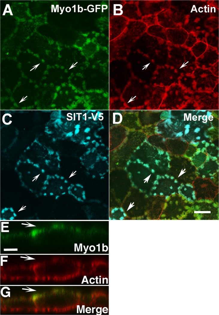
Fig 3. Expressed tagged rat Myo1b localized with the AATer SIT1 in apical microvilli of OK 3B/2 cells.
Myo1b-GFP (A, green) was found in patched microvilli, which are often located at the cell periphery of OK 3B/2 cells. The actin in the microvilli was identified by rhodamine phalloidin (B, red). SIT1-V5 (C, blue) localized to the apical microvilli with Myo1b. D is a merged image of A—C. Arrows identify actin-rich patched microvilli with both Myo1b-GFP and SIT1-V5. A-D are x-y images. Scale bar, A-D = 20 μm. In x-z sections (E-G), Myo1b-GFP (E, green) localized in OK 3B/2 cells primarily in actin-containing microvilli (F, red) on the apical plasma membrane. G is a merge of E and F, where yellow represents regions of overlap; the arrows point to the position of the apical microvilli in E-G. Scale bar, panels E-G = 10 μm.

