
Fig. 1.
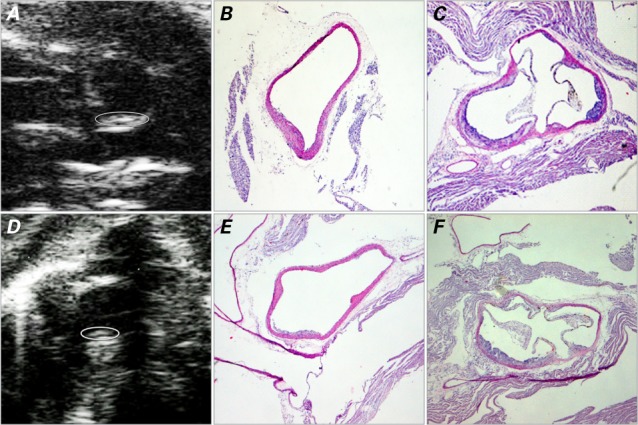
Images at 16 weeks show plaque development in the vascular lumen. In the IL-1Ra+/−/apoE−/− mice, the ascending aorta is shown on A) ultrasound biomicroscopy (UBM) (short-axis view) with B) the corresponding histologic image; C) histologic image of the aortic sinus (short-axis view). In the IL-1Ra+/+/apoE−/− mice, the ascending aorta is shown in D) UBM short-axis view with E) the corresponding histologic image; F) histologic image of the aortic sinus. The UBM images show the border of the plaque (circled) within the curvature of the ascending aorta. Histologic images are H & E stains, orig. ×4.
apoE = apolipoprotein E; IL-1Ra = interleukin-1 receptor antagonist