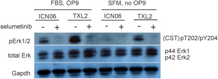
Fig 1. Western blot analysis shows pErk reduction through selumetinib treatment and by removal of stromal support.
ICNO6 and TXL2 ALL cells were cultured for 4 hrs in αMEM + 20% FBS on OP9 stroma or in αMEM + 1% BSA without OP9 stroma, and then treated in the same media for an additional 4 hours with 10 μM selumetinib. Western blots were incubated with the antibodies indicated in the panel. Gapdh, loading control. The membrane was sequentially stripped and re-probed with antibodies.

