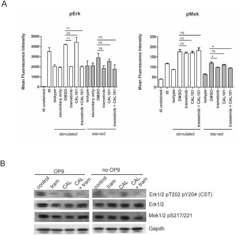
Fig 8. Mek pathway inhibition in combination-treated BCP-ALL cells by phospho-flow and Western blot.
(A) Phospho-flow on US7 cells using pErk1/2 (CST) or pMek (BD) antibodies in the left and right panels respectively, as indicated, in the presence or absence of trametinib, CAL101 or both. All samples were starved overnight in X-Vivo15 medium (t 0 samples), then treated from t 0 onward for 4 hours with αMEM + 20% FBS and OP9 stroma (‘stimulated’), or with αMEM + 1% BSA in the absence of stroma (‘starved’). At t 0 solvent DMSO or 10 μM of the indicated drugs was also added to the samples. Error bars, mean +/- SEM of three independent experiments. *p<0.05 **p<0.01, Student's t-test. (B) Western blot analysis (single analysis) of US7 cells grown for 24 hours in αMEM + 20% FBS and OP9 stroma (‘OP9’ samples, left panel), or in αMEM + 1% BSA in the absence of stroma (‘no OP9’ samples, right panel). Cells were then additionally treated for 4 hours with solvent DMSO (control) or 10 μM of the indicated drugs. Gapdh, loading control. Western blot membrane was sequentially stripped and reprobed with antibodies.

