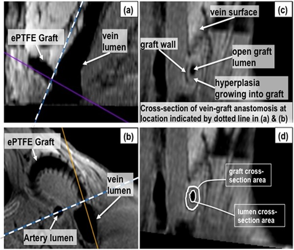
Fig 2. MRI analysis example.
A multiplanar reconstruction of the 2D black-blood scan was performed on a pig with a graft placed between the common carotid artery and external jugular vein in the neck. The sagittal plane (a) and transverse plane (b) of the vein-graft anastomosis region were used to locate the cross-section with the smallest open lumen area at the vein-graft anastomosis (c). The graft wall and vessel lumens appear hypointense (black) in black-blood imaging. The spiral support around the graft is also hypointense and visible along the graft wall in (b). The face of the plane indicated by the dashed line in (a) and (b) is shown in (c). The lumen and graft cross-sectional areas were measured as shown in (d) using the region-of-interest pencil tool in Osirix.

