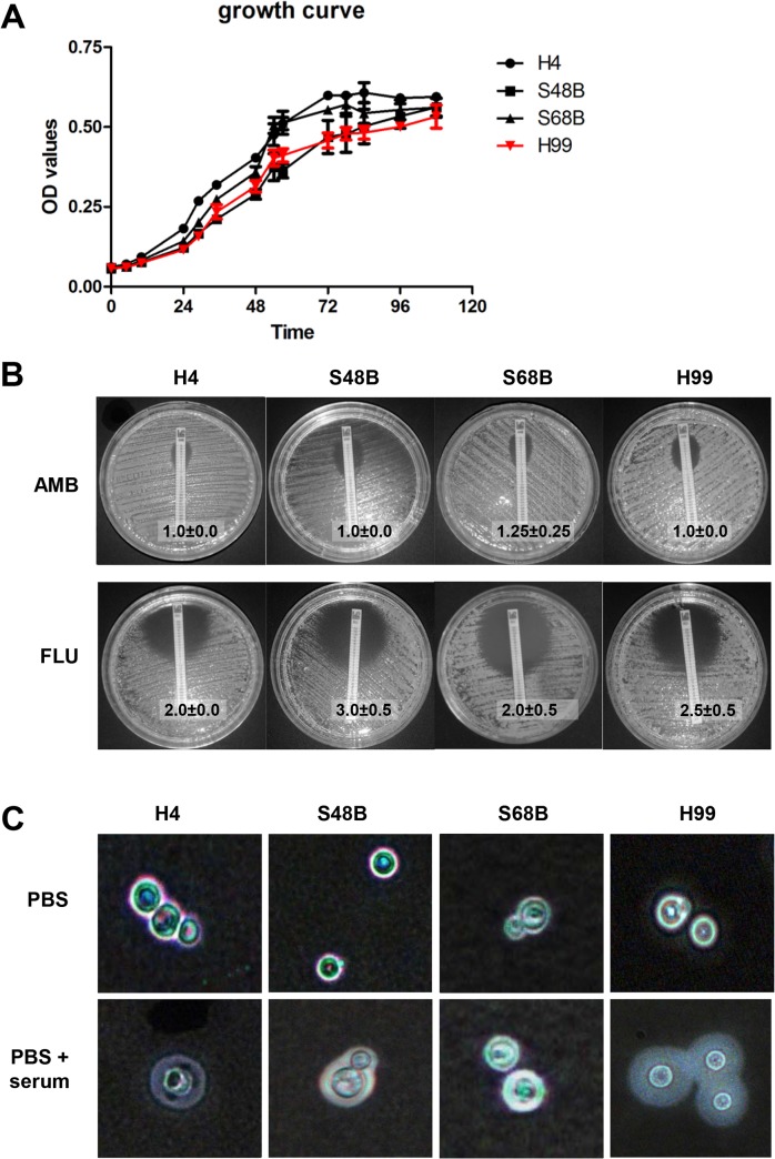
Fig 2. Different traits of C. neoformans strains.
(A) Growth curve. C. neoformans strains (H99, H4, S48B and S68B) cells were cultured in triplicates at 37°C with gentle shaking. OD reading for each sample was measured throughout a time course of 108 hours. (B) E-test. Fungal strains were inoculated evenly onto SDA plate. An AMB or FLU-containing MIC strip was then placed on each plate and incubated at 37°C for 48 hours. Eclipse sizes which intersected with the MIC strips were recorded. Shown were mean±SD from 2 duplicate plates. Data were representative of two independent experiments. (C) Capsule formation. Fungal cells were incubated in PBS with or without the presence of 10% serum, and incubated for 24 hours at 37°C. Cells were then applied on glass slide for India ink staining.

