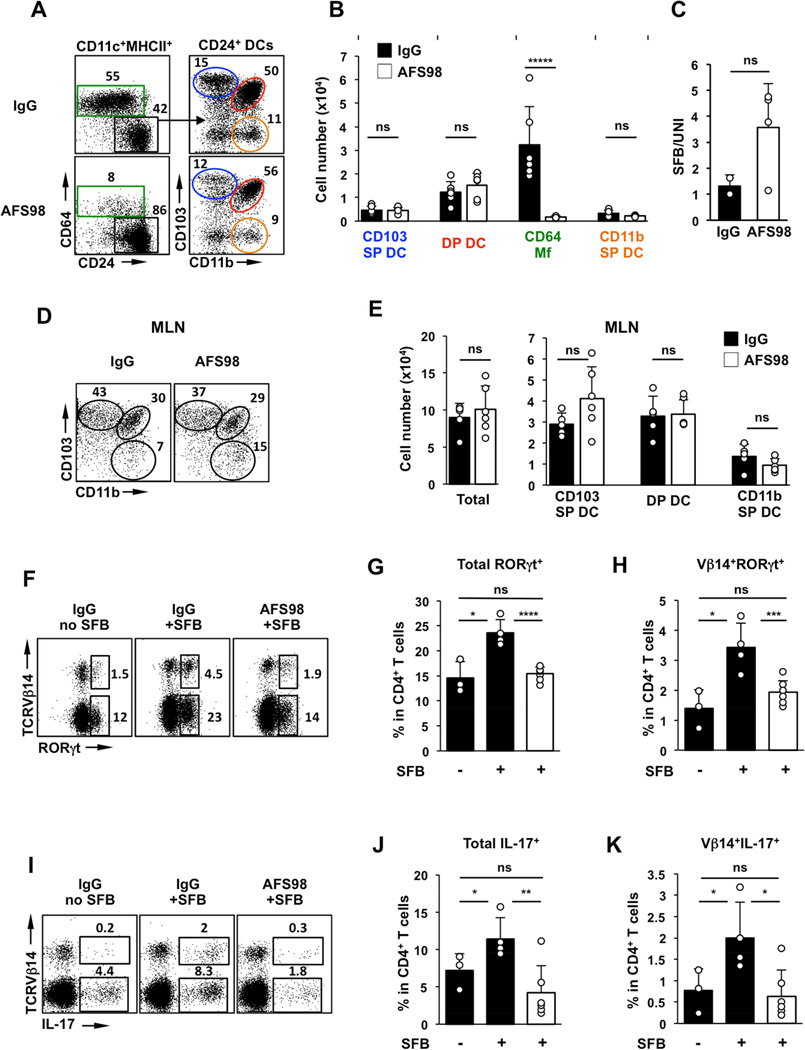
Figure 6. Treatment with CSF1R–blocking antibody impedes Th17 responses to SFB.
(A,B) MNP subsets in the SI LP of C57BL/6 mice treated with high dose of anti-CSF1R monoclonal antibody (AFS98) or control IgG four days before SFB colonization. (C) SFB levels in feces of AFS98 and control IgG treated mice normalized to total bacterial DNA (UNI). (D,E) Total cell numbers and cell numbers in MNP subsets in the migratory DC fraction of mesenteric lymph nodes (MLN). (F–H) RORγt+ Th17 cells in SI LP 8 days after SFB gavage. Plots gated on TCRβ+CD4+ cells. (I–K) IL-17+ Th17 cells in SI LP on Day 8 post SFB gavage. Plots gated on TCRβ+CD4+ cells.

