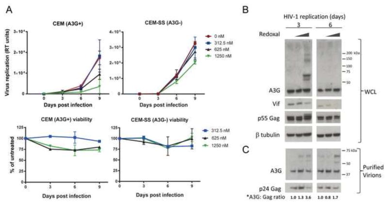
Figure 3. Redoxal inhibits HIV-1 replication in an A3G-expressing T cells.
A. HIV-1 spreading infection in redoxal-treated T cells. CEM and CEM-SS T cells were infected with HIV-1NL4-3 and 3 h post-infection cells were treated with 0, 312.5, 625, and 1250 nM redoxal. Virus production and cell viability of treated cells was tested at 3, 6, and 9 days post-infection by RT and CellTiter-Glo assays, respectively. HIV-1 replication is shown in RT units. Percentage of cell viability in redoxal-treated cells is relative to untreated controls (means of duplicate samples from independent wells ± SD). B. Redoxal induces the appearance of high molecular weight forms of A3G protein in HIV-1 infected CEM T cells. Infected cells treated with 0, 625, or 1250 nM redoxal were lysed 3 and 6 days post infection. Endogenous A3G, Vif, p55Gag, and β-tubulin protein levels in infected cell lysates were analyzed by Western blotting. WCL: Whole Cell Lysates. C. Redoxal induces the appearance of high molecular weight forms of A3G protein incorporated into HIV-1 virions. Virions collected 3 and 6 days post infection were normalized for equivalent RT units and purified through 20% sucrose. A3G and p24 Gag protein levels in virion lysates were detected by Western blotting. *Values shown below A3G and p24 Gag blots represent relative levels of virion-associated A3G protein in virions produced in HIV-1 infected cells treated with redoxal were determined by densitometry of bands using ImageJ software and normalization to untreated cells (DMSO control).

