
Figure 3.
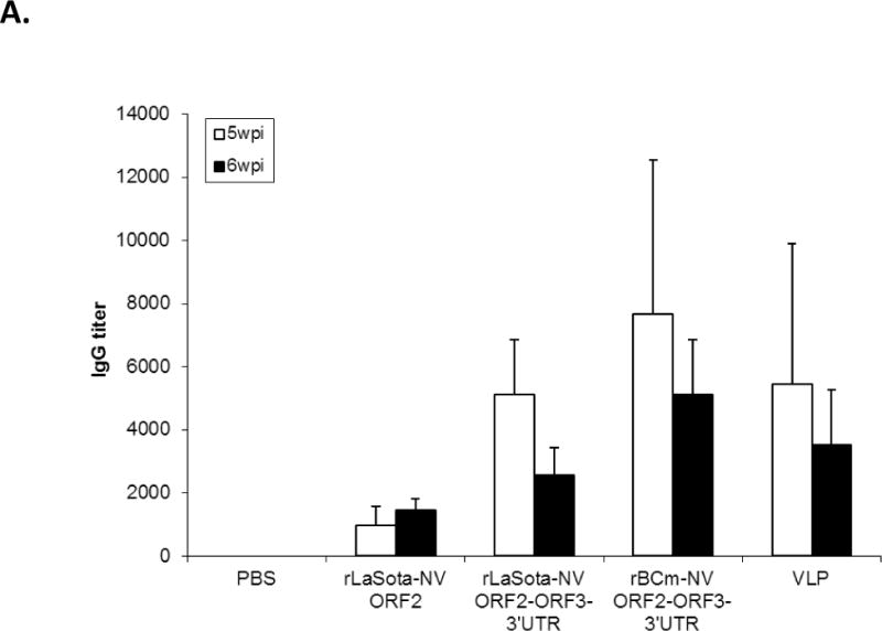
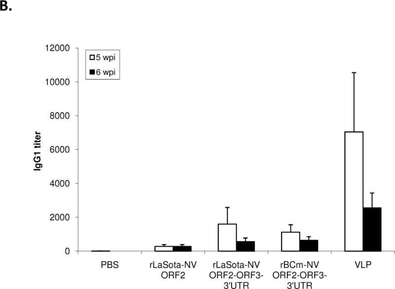
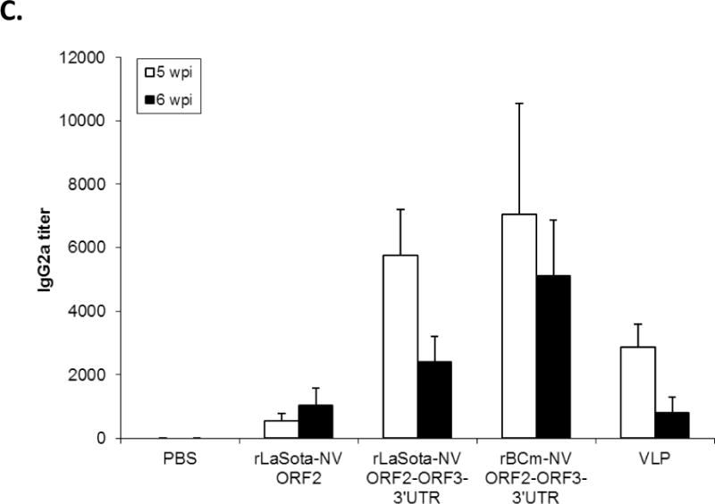
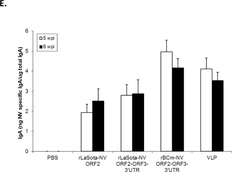
Antibody titers and NV-specific cellular and fecal IgA responses in mice after immunization with rNDVs and baculovirus-expressed VLPs. Mice were inoculated with each virus and VLPs by the intranasal route for three times in a two-week interval. (A) HBGA binding blocking assay was conducted to evaluate antibody-mediated neutralization for NV. Geometric mean titers (GMTs) for the values of the 50% blocking titer (BT50) determined by the H type 1 blocking assay. The titers of NV-specific total IgG (B) and subtypes IgG1 (C) and IgG2a (D) were determined by ELISA against purified baculovirus-expressed VLPs. The antibody titers were defined as the endpoint dilution with a cut off signal intensity of 0.2. (E) Splenocytes from the immunized mice were stimulated with NV VLP and analyzed for the production of IFN-γ, TNF-α, and IL-2 by the ELISPOT assay. The mean spot-forming cells (SFC)/106 cells with the error bars are shown. (F) Fecal samples were diluted in PBS, vortexed, and clarified by centrifugation. NV-specific and total IgA antibodies were determined by ELISA. The ratio between NV-specific IgA and total IgA was determined. Results are represented as mean ± sd for the mean of duplicate samples collected from 5 mice for each group. Statistical significance was determined by ANOVA (*P<0.05).