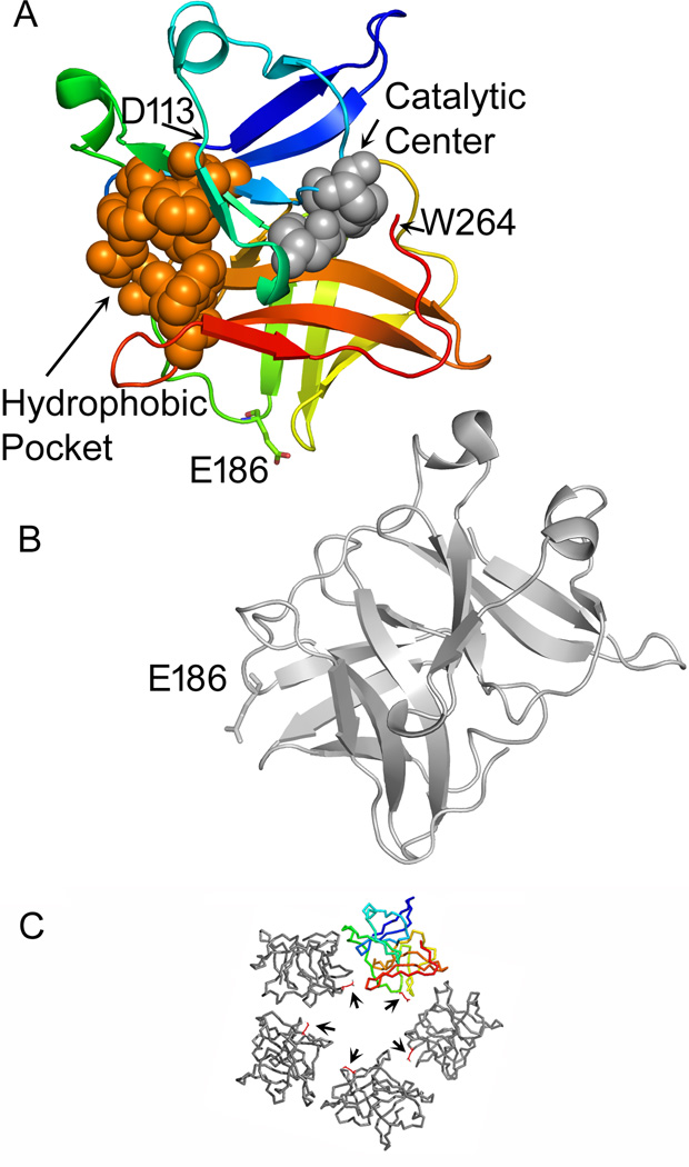
Figure 1. Location of E186 in the SINV capsid-capsid interface.
The fitting of the SINV capsid protease domain into the viral nucleocapsid structure is described in reference (Zhang et al., 2002) and (PDB: 1LD4). A capsid pentamer from that structure is shown in panel C, with E186 shown as a red stick structure (arrows). The capsid protein shown in color and the gray protein located clockwise to it in the pentamer in C are displayed in the larger cartoon structures in A and B respectively, with E186 indicated on each. The colored subunit (A) is displayed in blue (starting at the N-terminal D113) to red (C-terminal W264). The residues that form the catalytic triad (H141, D163, and S215) are shown as space filling structures in grey. The residues that form the hydrophobic pocket into which the E2 cytoplasmic tail inserts (Lee et al., 1996; Skoging et al., 1996) are shown as space filling structures in orange. Figure was generated using PyMOL (DeLano, 2002).

