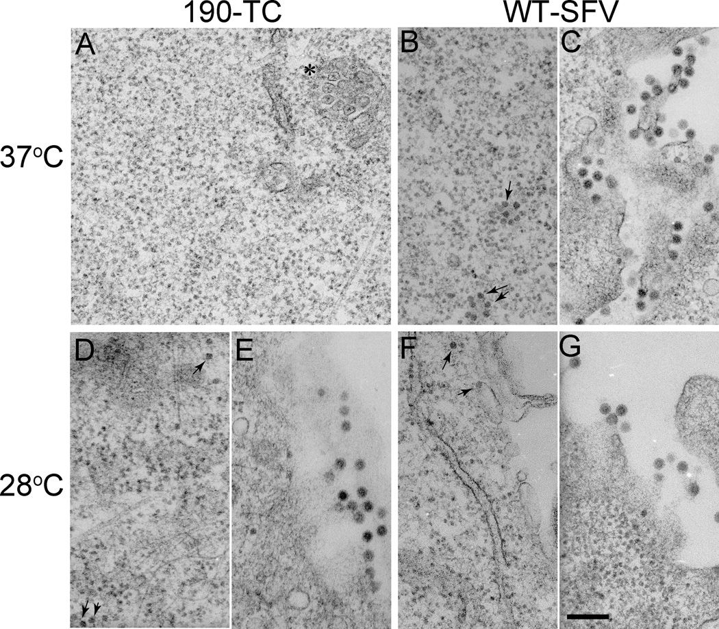
Figure 5. Electron microscopy of WT SFV or 190-TC mutant-infected cells.
BHK-21 cells were electroporated with WT SFV (B, C, F, G) or 190-TC mutant (A, D, E) viral RNA and incubated at 37°C for 2 h. Cells were then incubated at 37°C for 10 h (A, B, C) or 28°C for 18 h (D, E, F, G), and processed for electron microscopy. Panels C, E, and G show representative examples of the morphology of budding virus particles. Panel B, D, and F show representative examples of cytoplasmic nucleocapsids (indicated by arrows). Panel A is a representative image of 190-TC infected cells at 37°C with a cytopathic vacuole I structure indicated by the asterisk. All images were acquired at a magnification of 20,000 X, with the scale bar representing 200 nm.

