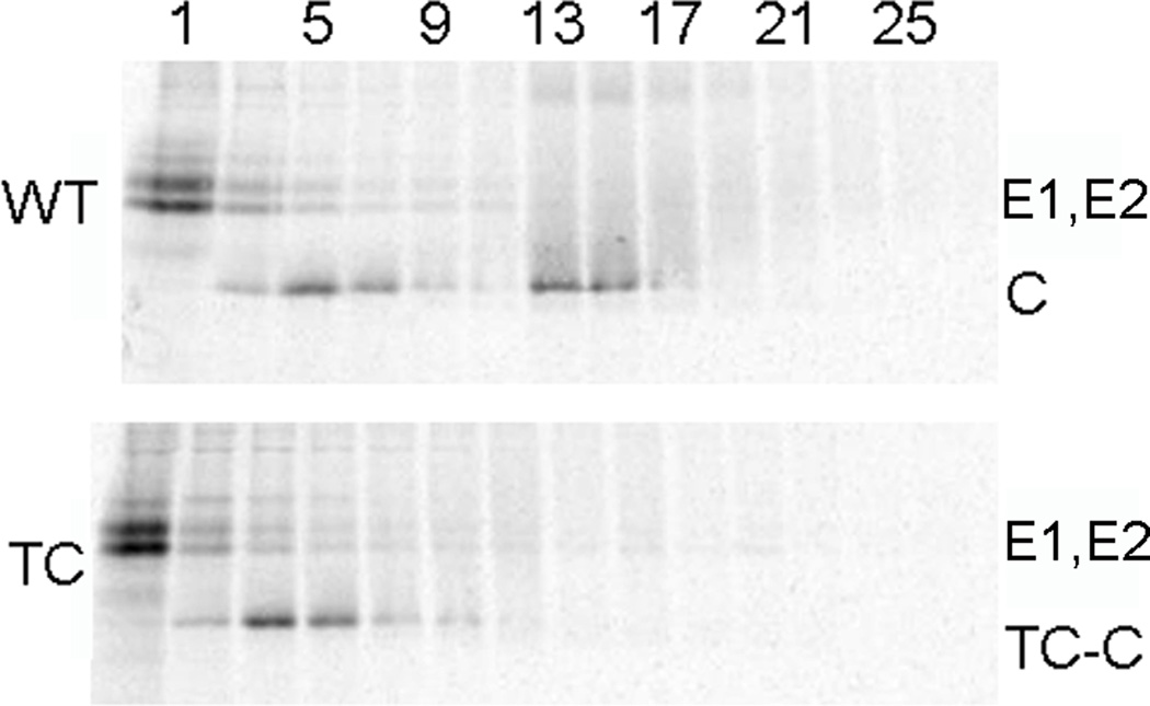
Figure 6. Gradient stability of 186-TC SINV cytoplasmic nucleocapsids.
BHK-21 cells were infected with WT or 186-TC mutant SINV and labeled for 14 h with [35S]-methionine/cysteine at 28°C. Cells were then solubilized in lysis buffer, layered onto sucrose gradients, and separated by sedimentation. Aliquots of the fractions were analyzed by SDS-PAGE, with fraction 1 representing the top of the gradient. The positions of the E1, E2 and capsid (C or TC-C) proteins are indicated. Shown is a representative example of three independent experiments.

