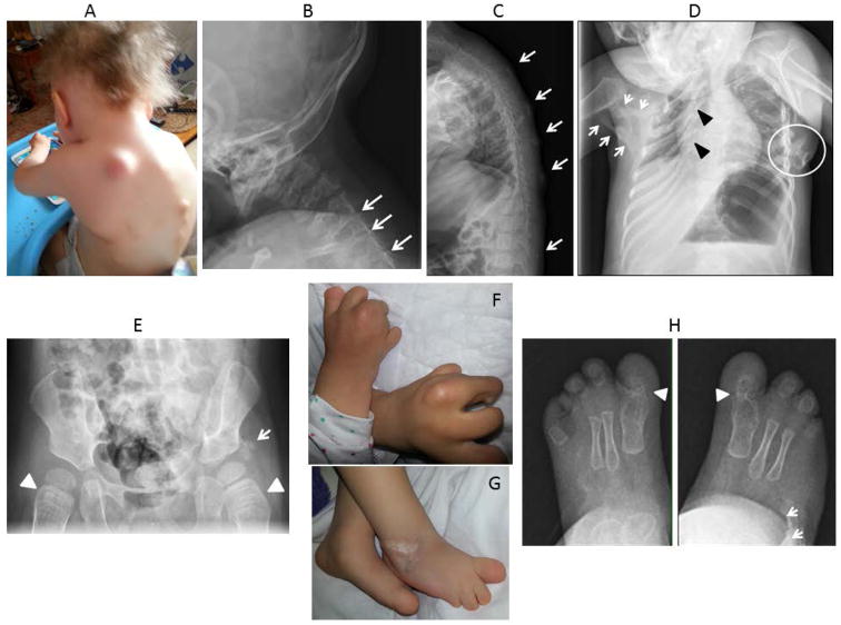
Figure 2.
Clinical and radiographic findings in Patient 2. A. Photograph of the neck and upper back at 30 months of age shows multiple FOP flare-ups as well as sparse scalp hair and severe upper limb malformations. B. Lateral radiograph of cervical spine at 13 months of age reveals orthotopic ankylosis of the posterior elements of C4-C5, C5-C6 and C6-C7 (arrows). C. Lateral radiograph of the thoracic spine at 22 months of age shows multiple areas of heterotopic ossification (arrows). D. Anteroposterior radiograph of the chest at 22 months of age shows severe right rib malformations (black arrowheads), severe restrictive heterotopic ossification of the right chest and shoulder girdle (white arrows) and osteochondromas of the left chest wall (white circle). E. Anteroposterior radiograph of the pelvis at 26 months of age shows short broad femoral necks (arrowheads) and heterotopic ossification adjacent to the left acetabulum (arrow). F. Photograph of hands shows severe malformations of all digits, reduction deficits and lack of nails. G. Photograph of feet shows severe malformations of all digits, reduction deficits and lack of nails. H. Anteroposterior radiographs of the feet at 13 months of age reveal bilateral malformations of the great toes with fusion and malformation of the first metatarso-phalangeal joint (arrowheads), evidence of symmetrical syndactyly, and heterotopic ossification of the right mid foot (arrows).

