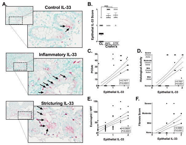
Figure 5. Epithelial-IL-33 expression is associated with disease progression and eosinophilia in complicated Crohn’s disease.
Representative photomicrographs of IL-33 immuohistochemically stained ileal tissues from (Ai) control (CL) subjects or from patients with (Aii) inflammatory (INFL) or (Aiii) stricturing (STR) CD. Arrows indicate epithelial-IL-33. (B) A score was generated to quantify the absence (0), presence (1) or increased presence (2) of epithelial-IL-33 staining. Epithelial-IL-33 scores were analyzed for relationships to subjects’ (C) pediatric Crohn’s disease activity index (PCDAI), (D) histological activity and (E) peak eosinophils / hpf (EPX) and (F) fibrosis score measured in H&E stained tissues. All analyses were performed on subjects defined in Table 1 that were not considered under treatment relevant to their CD. Statistical significance was assessed for epithelial-IL-33 scores by using 1-way ANOVA with Newman-Keuls multiple comparisons test. ***P≤0.001. For all other analysis the Pearson’s correlation coefficient (r) and its associated statistical significance are shown.

