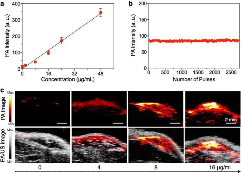
Figure 4. In vivo PA properties of SPN4.
(a) PA intensities of SPN4–matrigel inclusions (30 μL) in the subcutaneous dorsal space of living mice as a function of nanoparticle mass concentration. The tissue background signal, calculated as the average PA signal in areas where no nanoparticles were injected, was 25±6 a.u. R2 = 0.991. (b) PA amplitudes of SPN4–matrigel inclusion (16 μg/mL, 30 μL) in the subcutaneous dorsal space of living mice versus number of laser pulses. (c) PA (upper) and PA/ultrasound co-registered (lower) images of the nanoparticle–matrigel inclusions in mice at indicated concentrations. The images represent transverse slices through the subcutaneous inclusions. A single laser pulse at 750 nm with a laser fluence of 9 mJ/cm2 and a pulse repetition rate of 20 Hz was used for all experiments. Error bars represent standard deviations of three separate measurements.

