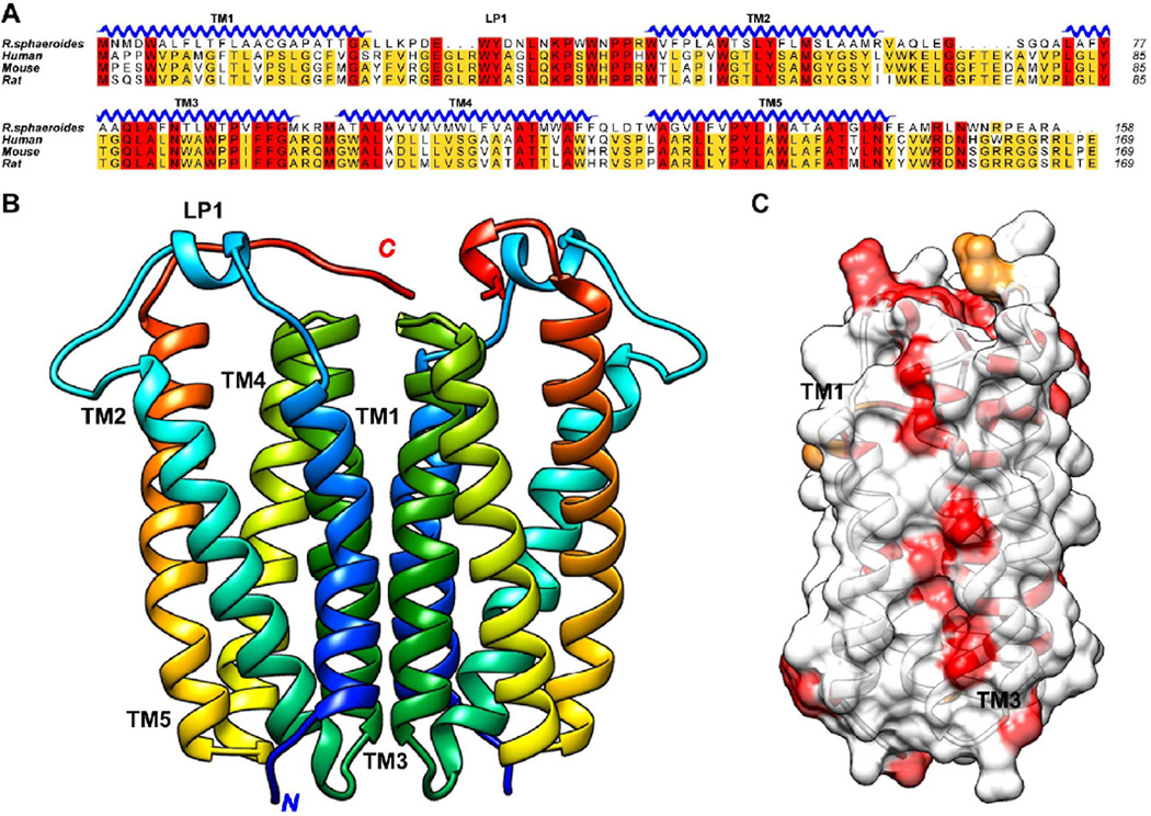
Figure 1. Dimeric structure of RsTSPO and the conservation of the dimer interface.
(A) Sequence alignment showing the most conserved residues in red, less conserved in yellow, unconserved in white. The alignment demonstrates that R. sphaeroides TSPO is closely related to its mammalian homologs. (B) RsTSPO crystal structure shows a dimer composed of two monomers of 5 transmembrane helices (TM), with TM-III (green) contributing most strongly to the interface interaction. (C) Highly conserved surface residues in TM-III at the dimer interface, indicated in red. (Made in Chimera from PDB#: 4UC1)

