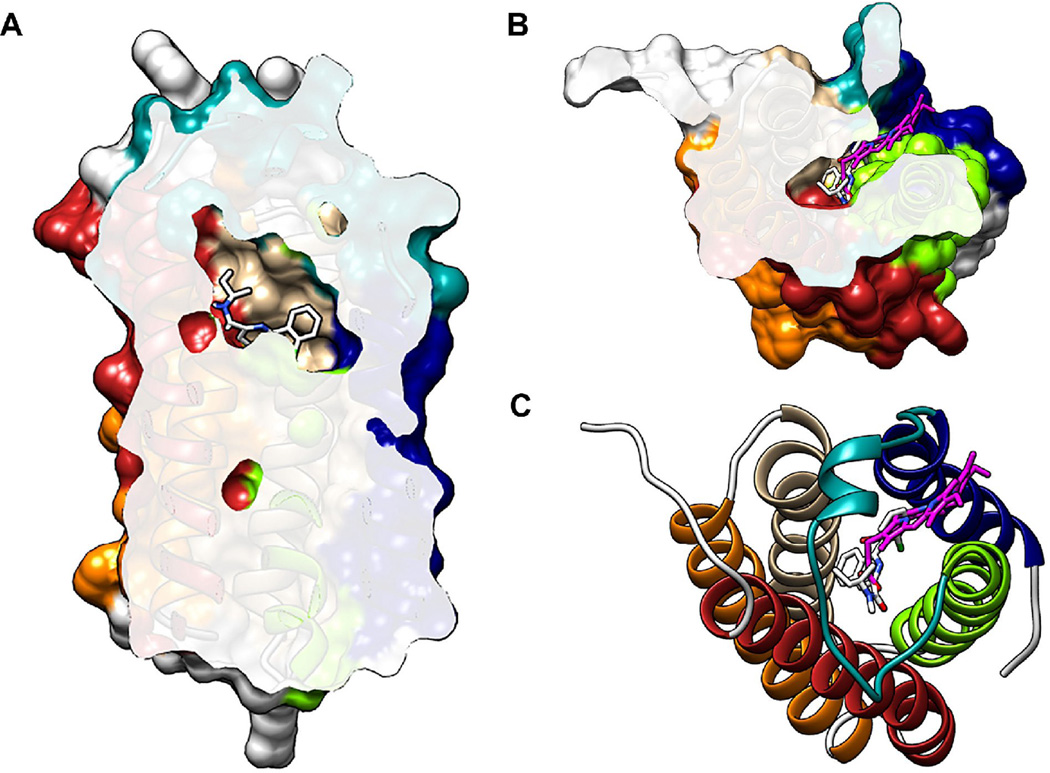
Figure 2. Ligand binding sites in the crystal structure of RsTSPO.
A highly similar central cavity is seen in the crystal structures of both Rhodobacter and Bacillus TSPO with a loop (teal) covering the top. (A) Cutaway of RsTSPO showing the central cavity with PK11195 (white) from BcTSPO modeled in the cavity. (B, C) The porphyrin (magenta) resolved in RsTSPO binds in the same cavity as PK11195, in a partially overlapping site. The protein surface is shown in A and B and a cartoon representation in C. (Figures were made in Chimera from PDB# 4UC1 and PDB# 4RY1.)

