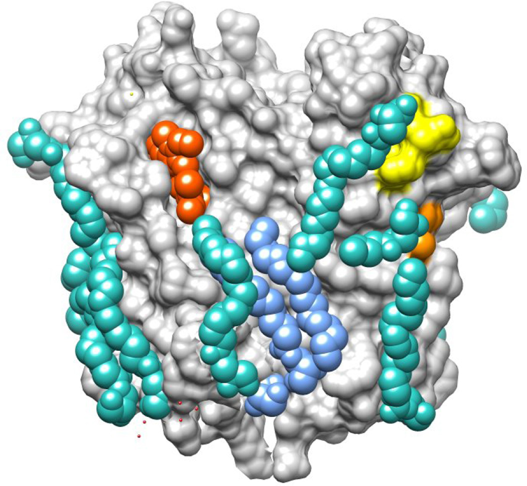
Figure 3. Crystal structure of RsTSPO with bound lipids and ligands.
RsTSPO crystal structure is shown in a surface rendering. Phospholipid is shown in light blue, monooleins in cyan, prophyrin in red, the CRAC site in yellow and the LAF site in orange. The human polymorphism A147T (A139T in RsTSPO) is located immediately above the LAF site. (Produced in Chimera from PDB# 4UC1.)

