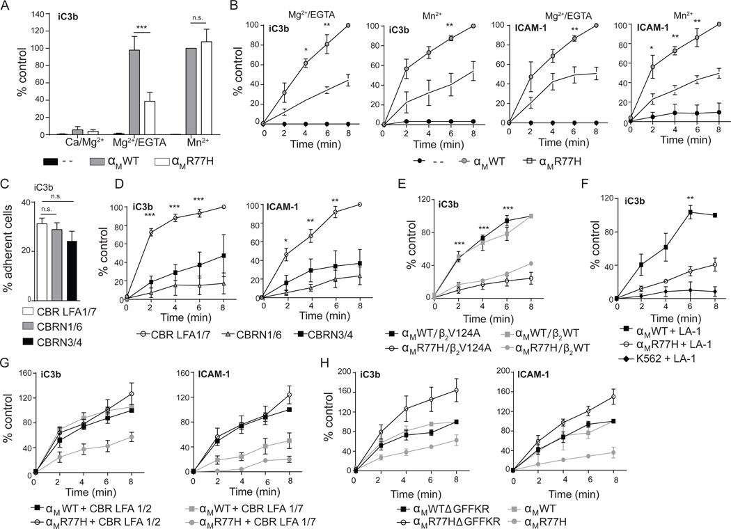
Figure 2. Functional effects of R77H in K562 cells.
K562 cells lacking β2 integrins (--), or stably expressing αMWT or αMR77H with β2 were evaluated for adhesion to iC3b and ICAM-1 following activation with Mn2+ or Mg2+/EGTA, under static conditions or flow conditions. (A) Adhesion of αMWT or αMR77H cells to iC3b-coated surfaces under static conditions. Results are presented as percent of adherent cells relative to cells expressing αMWT plus Mn2+. n=6 experiments. (B) Adhesion of αMWT or αMR77H cells to iC3b and ICAM-1 under shear flow (0.38 dynes/cm2) evaluated at the indicated time points. Results are presented as percent of adherent cells relative to cells expressing αMWT at 8min. n=3–4 independent experiments. (C–D) Binding of αMWT- cells to ligand-coated surfaces following treatment with indicated β-propeller antibodies (as in Figure 1) under static (C) or shear flow (D) conditions. (E) Cell adhesion under shear flow in cells expressing αMWT or αMR77H with β2WT or β2V124A. (F–G) Adhesion of αMWT or αMR77H cells pretreated with leukoadherin-1 (LA-1) or vehicle control (F) or CBR LFA1/2 or control antibody, CBR LFA1/7 (G). (H) Adhesion of cells expressing αMWT or αMR77H with or without a GFFKR deletion (Δ). n=3 for panels E–H. **p<0.01, ***p<0.001. See also Figure S2, S3 and Table S1.

