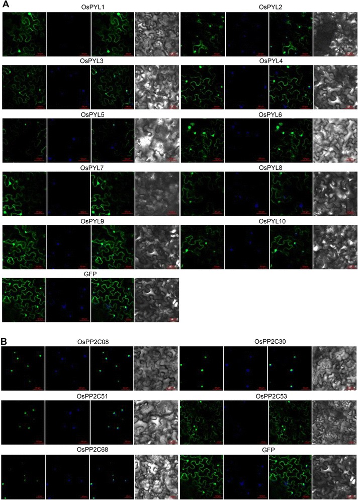
Fig. 2.

Subcellular localization of OsPYLs and OsPP2Cs. Confocal images were taken from Nicotiana benthamiana leaves epidermal cells. Constructs of (GFP)-OsPYLs (a) and (GFP)-OsPP2Cs (b) driven by the 35S promoter were infiltrated and observed at 3 days later. From left panel to right panel are GFP image, DAPI dye image, merged image and bright-field image. Empty GFP vector was used as control. The positions of nuclei were shown by DAPI staining
