
Figure 3.
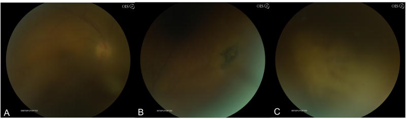
Fundus photograph of patient with HSV-2-ARN with moderate vitreous haze (A). Superonasally there was a patch of retinal pigment epithelial hyperpigmentation suggestive of toxoplasmosis or HSV-2 ARN, which characteristically shows pigmented chorioretinal scars (B). Inferonasally and nasally there are patches of confluent retinal whitening with hemorrhages and vasculitis (C).