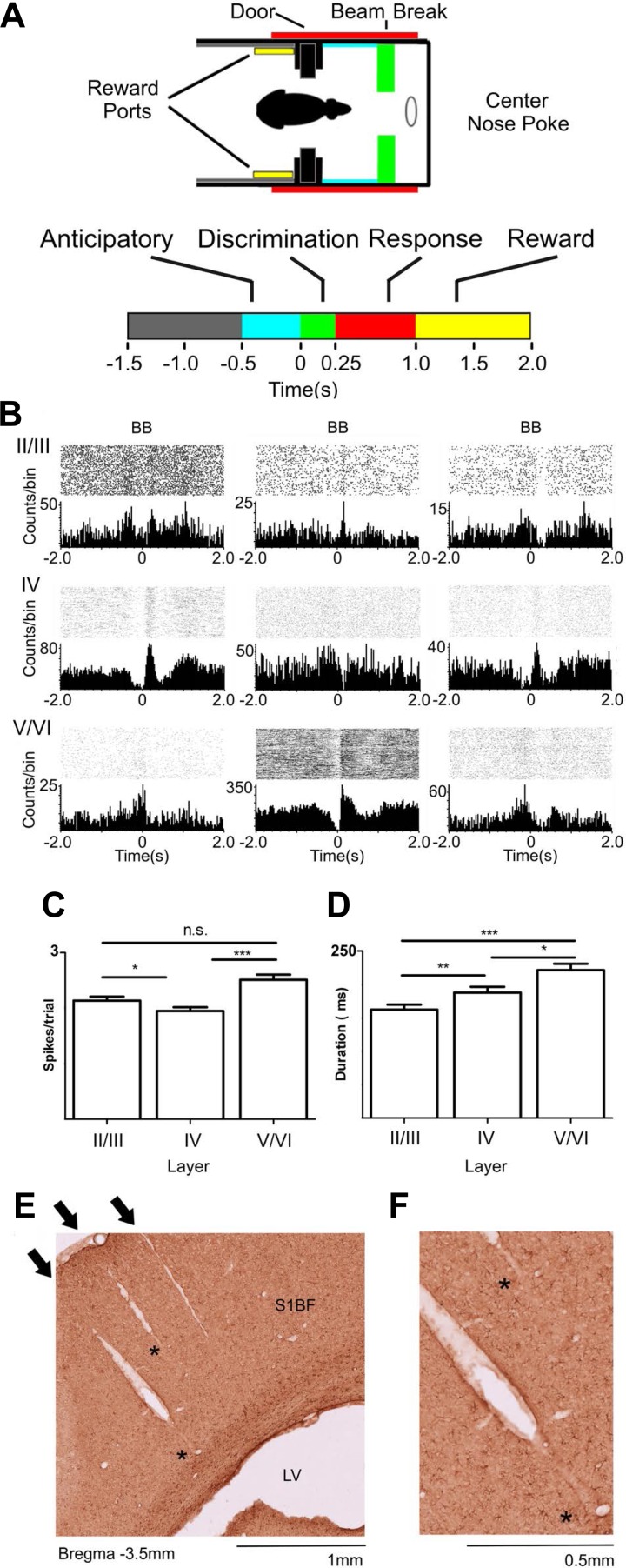
Fig. 1.
Layer-specific activity during tactile discrimination. A: active tactile discrimination task with different behavioral epochs, respectively: baseline (gray), anticipatory (cyan: −500-0 ms), discrimination (green: 0–250 ms), response (red: 250–1,000 ms), and reward (yellow: 1,000–2,000 ms) periods. B: perievent raster histograms from representative cells recorded from different depths in S1. BB, beam break in the discrimination bars (time = 0 s). Multiple different types of neuronal firing rate modulations were found. These neuronal firing modulations could be facilitated or suppressed and were present in all layers at all different periods of the task. C: layer-specific differences in magnitude of facilitated firing modulations during a trial. Neurons recorded from layers V/VI presented the largest neuronal firing rate modulations, followed by firing modulations recorded from layers II/III, and lastly by the ones recorded from layer IV. D: duration of neuronal firing modulations was layer specific during tactile discrimination. Neurons recorded from layers V/VI presented the longest facilitated firing modulations, followed by neurons recorded from layer IV, and lastly by neurons recorded from layers II/III. Error bars represent SE. *P < 0.05, **P <0.01, ***P < 0.001. E: example of 3 electrode tracks in the S1 cortex barrel field (S1BF). Note that the electrode tracks are orthogonal to the cortical surface and extend throughout multiple layers. The arrows indicate the point of entry in the cortical surface. The asterisks indicate places where the electrode tracks are present but not as clearly visible. LV, lateral ventricle. F: same histological preparation as E magnified to show the electrode tracks indicated by asterisks in E.

