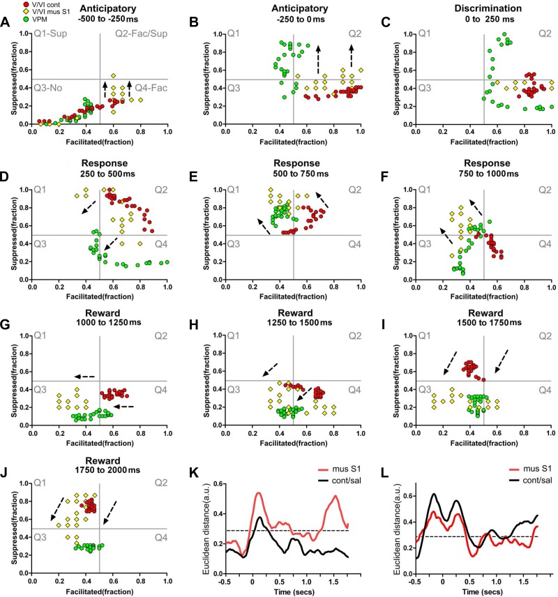
Fig. 11.
Bilateral integration is associated with paralemniscal neuronal dynamics in infragranular layers. Each colored circle or diamond depicts simultaneously the fraction of facilitated and suppressed neuronal firing rate modulations in a 10-ms bin. Red circles represent neuronal state maps of layers V/VI in control conditions, yellow diamonds represent neuronal state maps of layers V/VI after contralateral S1 inactivation, and green circles indicate neuronal state maps of VPM. The black intermittent lines indicate the overall translational shift observed in the neuronal state maps after S1 contralateral inactivation. A–J: neuronal state maps of layers V/VI after contralateral S1 inactivation presented multiple shifts that were now closer to the neuronal state maps of VPM, suggesting that bilateral S1 integration may function as a switch between preferential lemniscal or paralemniscal modes of processing. K: during control conditions (black line), the Euclidean distance between neuronal state maps of layers V/VI and POM was smaller than after S1 inactivation (compare black to red line). L: after S1 inactivation, the Euclidean distance between the neuronal state maps of layers V/VI of S1 and VPM became closer (compare black to red line). Overall the inactivation of S1 increased the Euclidean distance between neural state maps of infragranular layers and POM and reduced the Euclidean distance between the neural state maps of infragranular layers and VPM. Mus, muscimol.

