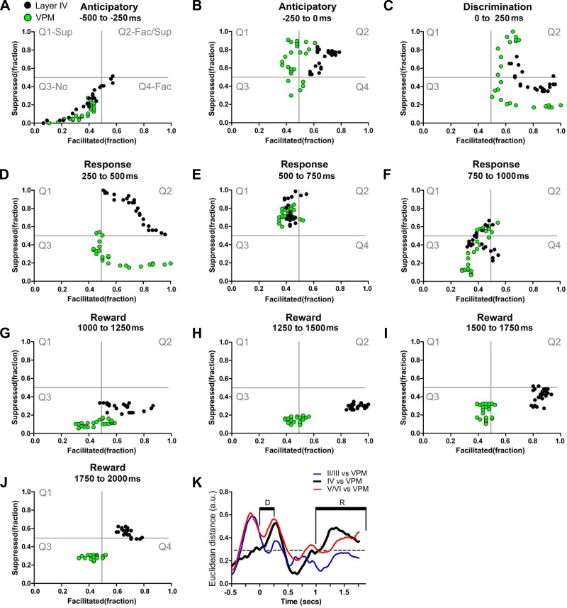
Fig. 5.
Ventral posterior medial nucleus of the thalamus (VPM) and granular layer share many dynamical neuronal modulations. Each colored circle depicts simultaneously the fraction of facilitated and suppressed neuronal firing rate modulations in a 10-ms bin. Neuronal state maps of layer IV (black dots) and VPM (green dots) are shown. A–J: neuronal state maps calculated from neurons recorded in VPM and layer IV of S1 were similar during several periods of the active tactile discrimination task. Major differences were found during the 1st part of the response period (250–500 ms) and during most of the reward period (1,250–2,000 ms). K: Euclidean distance between neuronal state maps of different S1 layers and neuronal state maps of VPM. As suggested by the trajectories in the state map, neuronal state maps of layer IV are closer to neuronal state maps of VPM (black line) during the anticipatory and part of the animal behavioral response period. Euclidean distances between neuronal state maps from layers II/III and VPM (blue line) were maximal during the anticipatory period. Euclidean distances between neuronal state maps from layers V/VI and VPM (red line) were maximal during anticipatory, discrimination, and part of the animal behavioral response periods.

