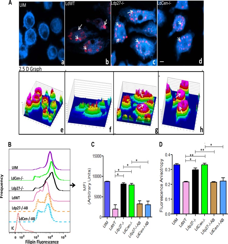
FIG 5.
Live attenuated parasite infection does not deplete membrane cholesterol and does not alter membrane fluidity of BMDM. BMDM were either left uninfected or infected with either RFP-tagged WT or RFP-tagged Ldp27−/− or mCherry-tagged LdCen−/− parasites for 24 h as described in Materials and Methods and then processed for confocal microscopy. (A) Cell preparations were stained with filipin and observed under a confocal laser-scanning microscope. The micrograph is representative of 3 independent experiments in which at least 100 cells per sample were analyzed. Bar, 10 μm. The corresponding filipin fluorescence intensity plots are shown below the respective micrographs, with the fluorescence intensity plotted on the z axis (a six-step rainbow look-up table was used to help visualize the range of intensity values within the micrographs). (B and C) In a separate experiment, BMDM were either left uninfected or infected with WT, live attenuated, or add-back parasites for 24 h. Macrophages were then stained with filipin and analyzed by flow cytometry. Control staining with isotype-matched antibodies was negative. Data are from 1 of 3 experiments conducted in the same way with similar results. *, P < 0.05; **, P < 0.005. (D) BMDM were either left uninfected or infected with various groups of parasites as described above. After 24 h, the membrane fluidity was estimated by calculating the fluorescence anisotropy value. The data represent the mean values ± standard deviations of results from 3 independent experiments that all yielded similar results. *, P < 0.05; **, P < 0.005.

