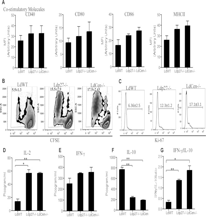
FIG 6.
Costimulatory molecule expression and T cell proliferation upon coculture of parasite-infected BMDM with OVA-specific transgenic T cells. (A) BMDM were infected with various groups of parasites as described in Materials and Methods for 24 h. The expression of MHC-II, CD40, CD80, and CD86 on BMDM was studied, and MFI were calculated based on flow cytometry results. (B) BMDM were pulsed with OVA peptide and infected with WT or live attenuated parasites for 24 h and then cocultured with purified CFSE-labeled CD4+ T cells from DO11.10 transgenic mice. CFSE dilution was measured in CD4+-gated T cells pulsed with different leishmanial lines. The experiment was independently repeated three times. (C) In a separate experiment, cell supernatants were collected from BMDM-CD4+ T cell coculture sets and stained with anti-Ki67 –PE and anti-CD4–FITC. T cell proliferation was estimated by flow cytometry by gating on Ki67+ CD4+ cells. Representative histograms shown here are from experiments repeated independently for three times. (D to F) Culture supernatants were collected after 5 days of coculture, and cytokines IL-2 (D), IFN-γ (E), and IL-10 (F) were measured using ELISA kits as per the manufacturer's instructions. (G) The IFN-γ/IL-10 ratio was determined. The data represent the mean values ± standard deviations of results from 3 independent experiments. *, P < 0.05; **, P < 0.005 compared to WT-infected BMDM.

