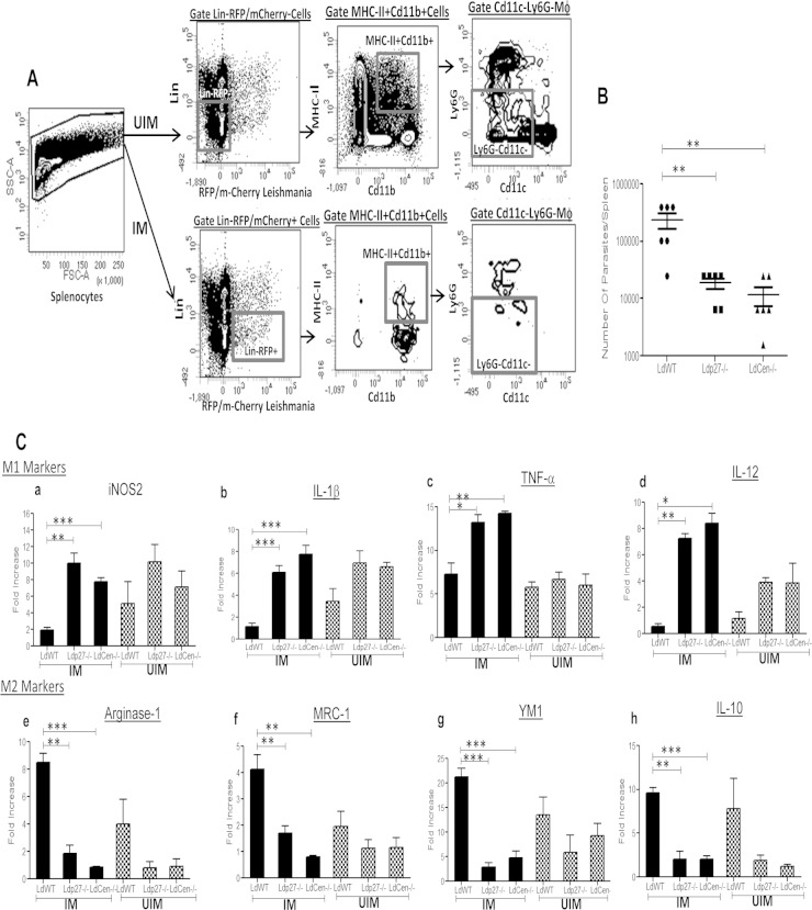
FIG 7.
Live attenuated parasite infection in mice induces classical activation (M1 phenotype) of parasitized macrophages isolated from spleens. (A) UIM were sorted from the spleens of different groups of mice (n = 6) by gating live single cells for lineage− (T cell, B cell, NK cell) RFP/mCherry– Cd11b+ MHCII+ Cd11c− Ly6G–) markers, whereas parasitized splenic macrophages (IM) were sorted by gating live single cells for lineage− (T cell, B cell, NK cell) RFP/mCherry+ Cd11b+ MHC-II+ Cd11c– Ly6G–) markers. The sorting strategy is displayed. The experiment was repeated 3 times with pooled digests from 6 spleens per experiment. (B) Parasite numbers in spleens of different groups of infected mice were measured 7 days postinfection. Means and standard errors of the means for six mice in each group are shown. Data are representative of two independent experiments. **, P < 0.005. (C) Sorted parasitized and uninfected macrophages from different groups of mice were subjected to RNA isolation as described in Material and Methods. Isolated total RNA was reverse transcribed and expression levels of different genes were analyzed as described in Material and Methods. Normalized expression levels of M1 markers, such as iNOS2, IL-1β, TNF-α, and IL-12, and M2 markers like Arg-1, MRC-1, YM1, and IL-10 were estimated. The data represent the mean values ± standard deviations of results from 3 independent experiments that all yielded similar results.*, P < 0.05; **, P < 0.005; ***, P < 0.0005 compared to WT-infected BMDM.

