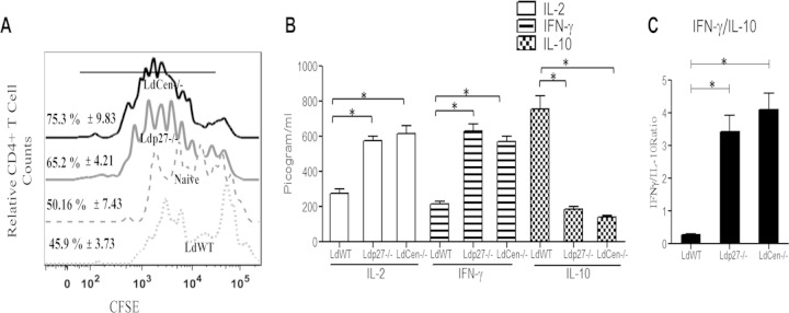
FIG 8.
BMDM from live attenuated parasite-infected mice are more efficient antigen-presenting cells than BMDM from WT-infected mice. BMDM isolated from BALB/c mice either uninfected or infected for 7 days with WT or live attenuated parasites were pulsed with OVA peptide and then cocultured with purified CFSE-labeled CD4+ T cells from DO11.10 transgenic mice. T cell proliferation was estimated by flow cytometry by studying CFSE dilution of gated CD4+ cells and is represented by the histogram. The staggered offset histogram overlay (A) displays the CD4+ T cell proliferation pattern as visualized by CFSE dilution after flow cytometry. (A) Cell proliferation was analyzed in triplicate experiments, and histograms representative of mean values were overlaid for the figure. The black bold line on the histogram overlay represents the percentage of proliferating CD4+ T cells. (B) The culture supernatant fluids were collected and assayed for IL-2, IFN-γ, and IL-10 in an ELISA. (C) The IFN-γ/IL-10 ratio was calculated from the data shown in panel B. The data represent the picogram levels of cytokines in culture supernatants and are presented as means ± standard deviations from three independent experiments. *, P < 0.05 compared to WT-infected BMDM.

