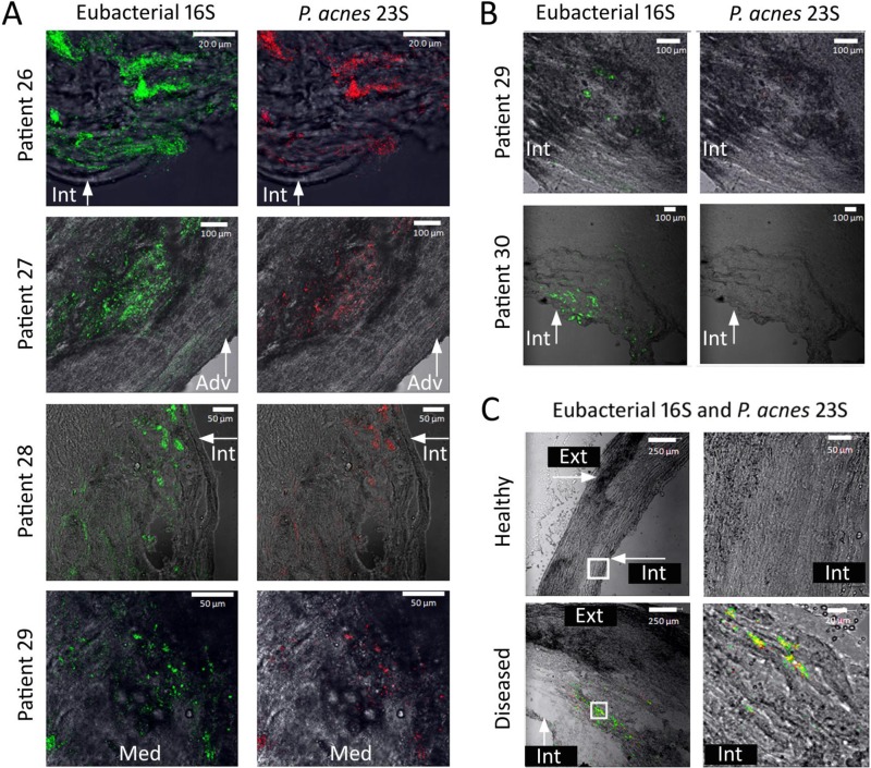
FIG 3.
Confocal laser scanning photomicrographs of FISH-probed atherosclerotic carotid arteries. (A) Locations and presence of eubacterial 16S rRNA gene- and P. acnes 23S rRNA gene-bound probes throughout atherosclerotic carotid arteries of 4 different patients. Green fluorescence indicates the presence of the bound eubacterial 16S rRNA gene probe, while red fluorescence indicates the presence of the bound P. acnes 23S rRNA gene probe. These samples contained both eubacterial 16S rRNA gene- and P. acnes 23S rRNA gene-bound probes. (B) Locations and presence of eubacterial 16S rRNA gene- and P. acnes 23S rRNA gene-bound probes throughout atherosclerotic carotid arteries of 2 different patients. Green fluorescence indicates the presence of the bound eubacterial 16S rRNA gene probe, while red fluorescence indicates the presence of the bound P. acnes 23S rRNA gene probe. These samples contained minimal to no detectable bound P. acnes 23S rRNA gene probe. (C) FISH-probed sample from patient 27, comparing a location of healthy tissue to a location where the atheroma was present. Locations examined under a higher magnification are indicated by white boxes. Green fluorescence indicates the presence of the bound eubacterial 16S rRNA gene probe, while red fluorescence indicates the presence of the bound P. acnes 23S rRNA gene probe. Healthy tissue contains no bound probe, while diseased tissue contains bound eubacterial 16S rRNA and P. acnes 23S rRNA gene probes. For all three panels, the anatomical location is indicated as follows: Int, intima; Med, media; and Adv, adventitia.

