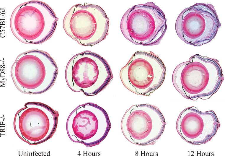
FIG 2.
Whole-eye histology of MyD88−/− and TRIF−/− eyes infected with B. cereus. C57BL/6J, MyD88−/−, and TRIF−/− mouse eyes were intravitreally injected with 100 CFU B. cereus. Uninfected and infected globes were harvested at 4, 8, and 12 h postinfection and processed for staining with hematoxylin and eosin. Uninfected C57BL/6J, MyD88−/−, and TRIF−/− eyes had no inflammation and normal retinal tissue structure. At 4 h, fibrin infiltration was observed in the anterior segment of C57BL/6J eyes, but retinal layers were normal. At 4 h, there was little to no fibrin infiltrate and retinal layers were normal in infected TRIF−/− and MyD88−/− eyes. At 8 and 12 h, infected C57BL/6J eyes had significant posterior segment inflammation and PMN infiltration, significant anterior and posterior segment fibrin accumulation, and disrupted retinal layers. In contrast, there was comparatively less fibrin and PMN infiltrates and retinal damage in infected TRIF−/− and MyD88−/− eyes at the same time points. Sections are representative of 4 eyes per time point with at least three independent experiments. Magnification, ×10.

