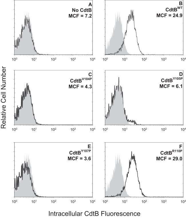
FIG 3.
Immunofluorescence analysis of internalization of CdtB CRAC site mutants in Jurkat cells. Jurkat cells were exposed to medium alone (gray curves), CdtA and CdtC alone (A), and CdtA and CdtC in the presence of CdtBWT (B), CdtBV104P (C), CdtBY105P (D), CdtBY107P (E), or CdtBR110P (F) for 1 h and then analyzed by immunofluorescence and flow cytometry for the presence of CdtB following fixation, permeabilization, and staining with anti-CdtB MAb conjugated to Alexa Fluor 488. Fluorescence is plotted versus relative cell number. Numbers represent the mean channel fluorescence (MCF). Note that the MCF for cells not exposed to any Cdt peptide was 5.7. At least 10,000 cells were analyzed per sample; results are representative of three experiments.

