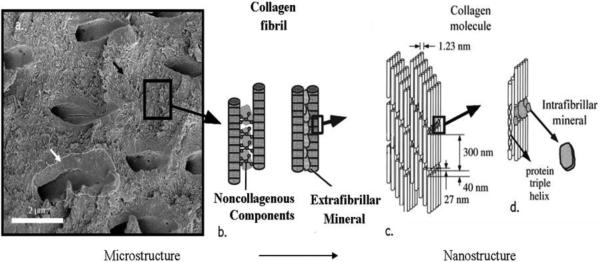
Fig.2.
(a) Scanning electron micrograph of a slice of dentin showing tubules, peritubular mineral (white arrow) and the collagenous intertubular matrix (black arrow); (b) Collagen fibrils interconnected by noncollagenous proteins and extrafibrillar mineral; (c) Collagen molecules display a typical spacing of 67 nm. Gap region equals 40 nm and the overlap region is 27 nm in length, which together gives the typical periodicity of 67 nm; (d) Intrafibrillar mineral particles are shown positioned in the gap region between collagen molecules. Reprinted with permission from Ref.7.

