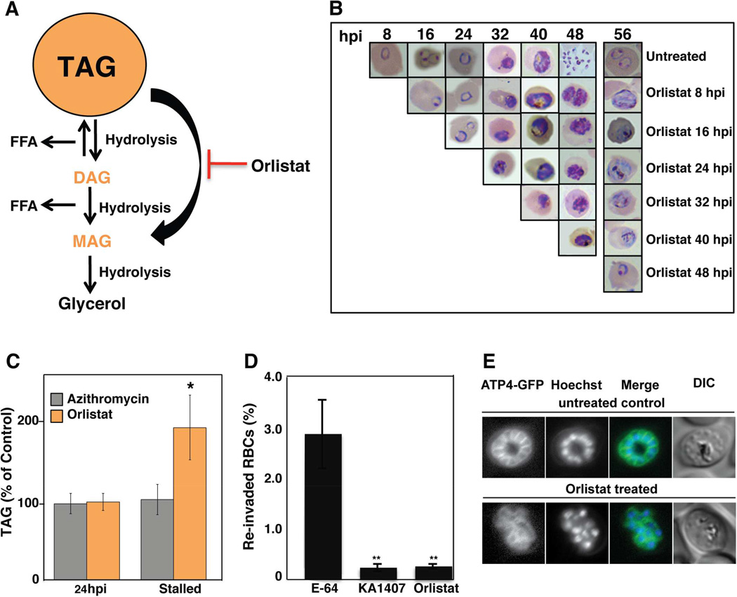
Figure 3. Orlistat Inhibits Triacylglycerol Degradation and Merozoite Maturation.
(A) Schematic representation of TAG degradation. TAG is stored in cytosolic lipid droplets (orange) and can be degraded by lipases to produce DAG or MAG. Orlistat prevents TAG catabolism by inhibiting lipase action. (B) Highly-synchronized ABS parasites (starting at 8 hpi) were serially assessed for development every 8 hr following the addition of 6 µM Orlistat (8×IC50), beginning at 8, 16, 24, 32, 40 and 48 hpi. Once added, Orlistat was maintained until the final time point at 56 hpi. Giemsa-stained smears were analyzed by light microscopy (examples provided) and the experiment was performed on three separate occasions. Across all experiments, parasites averaged 2.5–4% until reinvasion at 48 hr, when untreated parasites reinvaded and parasitemia climbed to 9–12%; however, reinvasion did not occur in Orlistat-treated parasites and parasitemias remained at 2–2.5%. (C) Synchronized ABS parasites were treated 6–8 hpi with [3H]-oleic acid (18:1) and 8×IC50 concentrations of either Orlistat or the delayed death inhibitor azithromycin. DMSO was used as a solvent control. Parasites were harvested at 24 and either 48 hr later (~56 hpi) for Orlistat or 72 hr later for azithromycin (corresponding to 30–32 hpi in the second cycle of growth), such that for both agents parasites had developmentally stalled at the later time points. Lipids were extracted and subjected to thin layer chromatography. TAG levels were normalized to DMSO-treated parasites extracted at the corresponding time points and are shown as means±SEM (n=3 independent experiments performed in triplicate). (D) Percent reinvasion of RBCs after mechanical rupture of synchronized schizont-infected RBCs treated with 1 µM E-64, 125 nM KAI407 or 6 µM (8×IC50) Orlistat. Data are shown as means±SEM (from three independent experiments performed in triplicate). (E) Plasma membrane ingression around developing daughter cells was visualized using the plasma membrane marker PfATP4-GFP (McNamara et al., 2013). Synchronized parasites were treated at 24 hpi with either 6 µM Orlistat or DMSO, and imaged 28 hr later (i.e. 52 hpi). Control parasites had organized and fully enclosed developing merozoites, whereas plasma membrane ingression was perturbed by Orlistat treatment. Nuclei were stained with Hoechst 33342.

