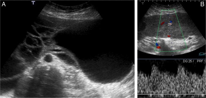
Figure 1.

Uterine subserosal leiomyoma with diffuse hydropic degeneration in a 35-year-old woman. Transabdominal ultrasound image in B mode (A); colour and spectral Doppler ultrasound image (B). A giant, well-defined, multilobulated abdominopelvic mass, with prominent vascularised solid as well as cystic components, is seen (A). Note the prominent vessels within the mass, clearly depicted by colour and spectral Doppler ultrasound imaging (B).
