Fig. 5.

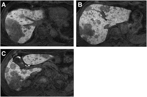
Abdominal contrast-enhanced magnetic resonance imaging before the second tumor biopsy. a–c Some tumors show heterointensity, and other tumors show central enhancement in the parenchymal phase of T1-weighted image

Abdominal contrast-enhanced magnetic resonance imaging before the second tumor biopsy. a–c Some tumors show heterointensity, and other tumors show central enhancement in the parenchymal phase of T1-weighted image