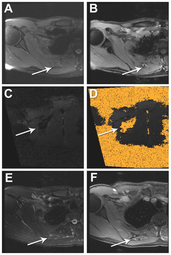
Figure 3. MRI-guided laser ablation of a slow-flow vascular malformation in the soft tissue between the right rhomboid and subscapularis muscles.
(A, B) Pre-ablation (A) axial T2-weighted and (B) gadolinium-enhanced axial T1-weight spoiled gradient echo (SPGR) MRI demonstrates heterogeneous abnormal T2 signal (A, white arrow) and contrast enhancement (B, white arrow) within the small vascular anomaly. (C, D) Intra-procedural coronal oblique (C) phase imaging demonstrates real-time tissue heating using proton resonance frequency MR thermometry and (D) thermal damage map calculated from this using the Arrhenius equation to estimate the ablation zone. (E, F) One-year post-ablation (E) axial T2-weighted MRI and (F) gadolinium-enhanced axial T1-weight spoiled gradient echo (SPGR) MRI demonstrates no significant T2 signal (E, white arrow) and no enhancement of the vascular anomaly (F, white arrow).

