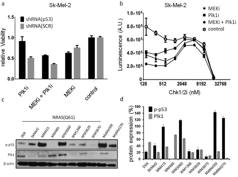
Figure 5. p-53 signaling affects Plk1 and MEK/Plk1 inhibitor efficacy.
a. Stable knockdown of p53 reduced the activity of Plk1 and MEK/Plk1 inhibition. p53 interference had no effect on viability in cells treated with the MEKi only, or on vehicle treated cells. (b) Additional Inhibition of Chk1/2 markedly reduced viability in MEK/Plk1 treated compared to single inhibitor or vehicle treated controls in a biphasic manner. (c) Immunoblot analyses of p-p53 and Plk1 in all 10 NRAS mutant cell lines used in this study. All cells, except MM485, express p-p53. Plk1 protein expression was pronounced in NRAS(Q61) mutant cells. (d) Relative expression of the respective proteins compared to β/actin. (N=3; mean±SD; SCR=scramble control shRNA; MEKi(JTP-74057)=20nM; Plk1i(BI6727)=100nM; Chk1/2i=PF-0477736)

