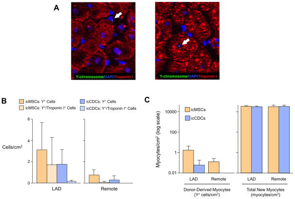
Figure 7. The Majority of New Myocytes Derived from Global icMSCs and icCDCs Arise From Endogenous Sources.
(A) Y-chromosomes were detected by Y-FISH. Y-FISH positive cells were primarily found in the interstitial space (left panel) with rare examples of Y+ cardiac myocytes (right panel). (B) The frequency of Y+ cells was low but similar in icMSC-treated and icCDC-treated animals. Rare Y+ cells also expressed Troponin I (hatched bars), indicative of cardiomyocyte differentiation. (C) The number of new myocytes differentiating from icMSCs and icCDCs was very small in comparison to the estimated increase in myocytes based on increases in myocyte nuclear density, implicating endogenous recipient cells as the primary source of new myocyte formation.

