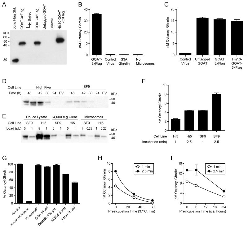
Figure 1. GOAT octanoylation assay establishment.
(A) Anti-Flag immunoblot of SF9 cells expressing various tagged GOAT constructs. As previously reported with GOAT expressed in human cells, boiling (10 min at 100°C) caused aggregation of GOAT and loss of signal. Control cells are infected with virus made from empty vector.
(B) GOAT octanoylation assay. Each 50 μl reaction was incubated for 5 min at 37°C with 50 μg microsome protein, 1 μM octanoyl-CoA, 50 μM palmitoyl-CoA, 10 μM Ghrelin27. Microsomes made with control virus made from empty vector and Ghrelin27-S3A are shown as controls.
(C) Activity of microsomes containing untagged GOAT, N-GOAT-3xFlag-C, and N-His10-GOAT-3xFlag-C under the same conditions as (B).
(D) Anti-Flag immunoblot of High Five and SF9 cells infected with GOAT-3xFlag virus for the indicated times. EV=virus made with empty vector, 48 hours. Each lane contains the equivalent of 20 μl suspension culture.
(E) Microsome preparation from 1 L cultures of SF9 and High Five (Hi5) cells. Loading shows two different amounts at each step and an equivalent fraction of the total is shown at each step.
(F) 25 μg microsome protein from High Five or SF9 cells were incubated for the indicated time at 37°C with 1 μM octanoyl-CoA, 50 μM palmitoyl-CoA, 10 μM Ghrelin27.
(G) The indicated protease inhibitors were pre-incubated with GOAT microsomes for 5 min before 1 min assay. PI cocktail: 2 μg/mL Leupeptin, Aprotinin, Pepstatin A, 2mM EGTA.
(H, I) GOAT microsomes stability. Microsomes were pre-incubated at the 37°C or on ice, respectively, at 95% of final assay concentration for the indicated times and then assayed for 1 or 2.5 min.

