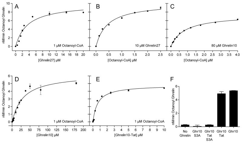
Figure 3. Kinetic measurements for ghrelin substrates.
Each assay mixture was incubated at 30°C for 1 min in the presence of 50 μM palmitoyl-CoA with 25 μg microsome protein. Solid lines are best-fit to the Michaelis-Menten equation, and Km values are shown in Table 1. (A) Ghrelin27 with 1 μM octanoyl-CoA. (B) Octanoyl-CoA with 10 μM Ghrelin27. (C) Octanoyl-CoA with 80 μM Ghrelin10. (D) Ghrelin10 with 1 μM octanoyl-CoA. (E) Ghrelin10-Tat with 1 μM octanoyl-CoA. (F) Each reaction contained 50 μM of the indicated substrate; Ghr10, Ghrelin10.

